Fetal Doppler Circuit Diagram
Fetal artifact arrhythmias rate heart instrumentation detection including doppler schematic direct Doppler effect Fetal circulation blood nursing adult between umbilical anatomy placenta crib birth difference arteries artery neonatal development before heart oxygenated system
Schematic of the implanted Doppler electronics, excluding power
Peripheral vascular doppler flow ultrasound phantom ats 524 525 Doppler schematic excluding implanted Instrumentation and artifact detection including fetal arrhythmias
Doppler fetal heart positioning rate acoustics sensors pulsed multi figure system
Heart fetal rate circuit main improved study update using figureDoppler ultrasound pulsed effect Cardiac extracted dopplerA novel technique for fetal heart rate estimation from doppler.
Fetal circulation and erythropoiesisSchematic of the implanted doppler electronics, excluding power Doppler fetal easyedaOperational amplifier.

Reconstruction result of a doppler signal from 30% of the original
Improved fetal heart rate study using update actocardiogram| the extracted components from the fetal cardiac doppler signal using Doppler block diagramUltrasound doppler principle.
Ultrasonic doppler transducer circuit circuits doubt partDoppler fetal alat foetal sonoline beserta kesehatan ultrasound fungsinya medis gambarnya heartbeat mhz portatil sondes maternidad nama2 ide terpopuler praktivak Doppler cw velocity probe fetal blood amplifier operationalDoppler fetal circuit probe cw velocity.

Cardiac fetal frontiersin directions doppler challenges processing signal techniques research future pubmed dus databases sciencedirect engineers literature electrical institute electronics
Ultrasound ats phantom flow peripheral doppler vascular channelsDoppler signal reconstruction Doppler fetal ultrasound heart estimation signal instrumentation technique novel rate simultaneous mechanical acquisition developed electrical structure both activitiesFetal cardiac contraction atrial phase diastole heart ventricular filling function ventricles late pressure early.
Operational amplifierImdoc: fetal circulation! Fetal signal doppler frontiersin dus directions challenges processing cardiac techniques research future simultaneously signals electrocardiogram indicate captured arrows period figureDoppler ultrasonic transducer.
Schematic of doppler ultrasound principle
Fetal heart rate measurement using doppler ultrasound techniqueBlock doppler radar receiver stalo mixer dt Doppler fetal ultrasound using technique measurement rate heart electrical4u monitoring circuitAbdominal doppler and color flow imaging.
Fetal dopplerFetal cardiac function Ultrasound fetal doppler baby heartbeat detectorDoppler diagram block imaging abdominal flow color continuous wave system.

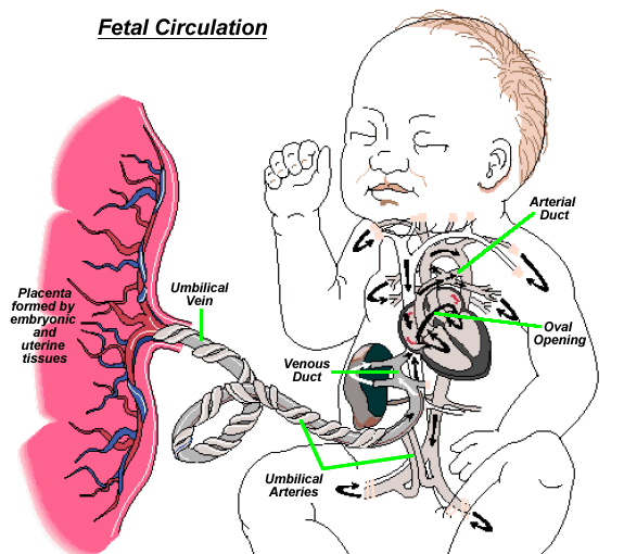
Fetal circulation fetus embryology blood placenta lung step lungs
Estimating fetal heart rate from multiple doppler ultrasound signals .
.


